Newsletter
By submitting this form,you consent to receive marketing updates from Durastab Medtech (Durastab IVD) .
Office Location
Shenzhen, China
Production Address
B1501, Meixun Science Park,
No.19, Jinxiu Zhonglu ,
Pingshan.
Durastab Medtech Co., Ltd. has always adhered to the mission of "making diagnostic reagents more stable and more accurate" since its establishment, and is committed to providing excellent ready-to-use reagent solutions to global advanced medical diagnostic companies.
To date, Durastab has successfully provided products to over 600 companies and research teams at thousands of universities and scientific research institutions worldwide, among which are more than 50 listed IVD companies.
About UsHRP Blotting Femto ECL is an ultra‑sensitive enhanced chemiluminescent substrate designed for low femtogram‑level protein detection in Western blotting. Compared to other commercially available substrates, this product delivers exceptionally high sensitivity and an excellent signal‑to‑noise ratio.
Tell us what you need and we will send the file to your email shortly.
HRP Blotting Femto ECL
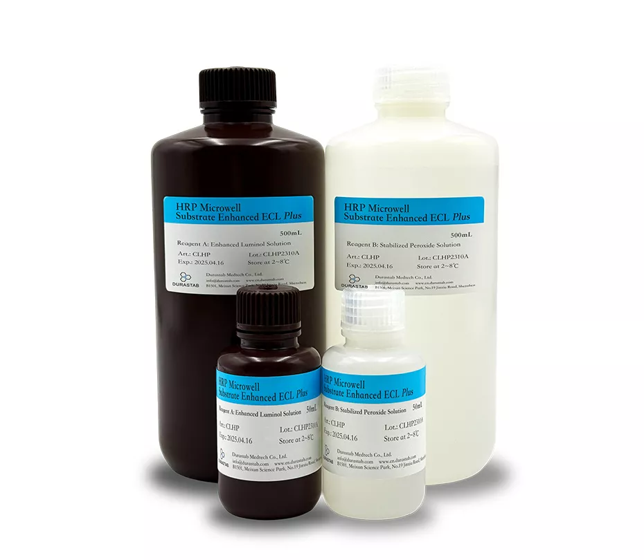
HRP Blotting Femto ECL is an ultra‑sensitive enhanced chemiluminescent substrate designed for low femtogram‑level protein detection in Western blotting. Compared to other commercially available substrates, this product delivers exceptionally high sensitivity and an excellent signal‑to‑noise ratio.
Art no: ECMF
Package size: 100mL, 1000 mL, bulk
Components:
Reagent A: Art no: ECMF-A, Enhanced Luminol Solution
Reagent B: Art no: ECMF-B, Stabilized Peroxide Solution
Shelf life: 3 years
Storage: 2-8℃ protected from light.
Application:
Western Blot: Use after 1:1 premixing
Characteristics:
① High Sensitivity: Low femtogram.
② Stability: Working solution remains stable for up to 8 hours at 4 °C.
③ Compatibility: Suitable for both nitrocellulose and PVDF membranes.
④ Extended Signal Duration: Signal persists for up to 8 hours.
⑤ Primary Antibody Dilution: 1:5,000–1:100,000 (mass concentration: 10–200 ng/mL).
⑥ Secondary Antibody Dilution: 1:100,000–1:500,000 (mass concentration: 2–10 ng/mL).
Instruction:
Western Blot Detection Using the Substrate:
1. Membrane Washing:
After protein transfer, remove the blot from the transfer apparatus and wash the membrane with deionized water for 5 minutes to remove residual transfer buffer.
2. Membrane Blocking:
Block the membrane with DuraBlot™ Western Blot Blocking Solution (e.g., 1× Casein Blocking Buffer, 1× Protein‑Free Blocking Buffer) at room temperature with shaking for 30–60 minutes, or alternatively, block statically overnight at 2–8 °C.
3. Primary Antibody Incubation:
Dilute the primary antibody to 10-200 µg/mL (or according to the manufacturer’s recommended dilution) using primary antibody diluent (or blocking solution). Incubate with the membrane with shaking for 1 hour at room temperature, or overnight at 2–8 °C.
4. Membrane Washing:
Wash the membrane with washing buffer (TBST, PBST, or other buffer containing 0.05% Tween 20) for 10 minutes. Repeat the wash twice.
5. Enzyme‑Labeled Secondary Antibody Incubation:
Dilute the enzyme‑labeled secondary antibody to 2–10 ng/mL or 1:100,000–1:500,000 using secondary antibody diluent. Incubate the blot with shaking for 30–60 minutes at room temperature.
6. Membrane Washing:
Wash the membrane 6 times with washing buffer, 5 minutes each wash, to thoroughly remove any unbound conjugated antibody.
7. Substrate Preparation:
Prepare the substrate working solution by mixing equal volumes of Substrate A (Enhanced Luminol Solution) and Stable Peroxide Solution B (e.g., 5 mL + 5 mL). Use sufficient volume to ensure complete wetting of the membrane without drying (0.1 mL/cm²).
8. Substrate Incubation:
Incubate the membrane with the substrate working solution for 5 minutes.
9. Membrane Imaging:
① Remove the blot from the working solution and place it in a plastic protector or clear plastic wrap.
② Remove excess liquid with lint‑free absorbent paper and carefully eliminate air bubbles from the membrane surface.
③ Image the blot using an imaging system.
Note: This product is highly sensitive for detection. Careful optimization of individual assay components (antibodies, conjugates, etc.) is essential to minimize nonspecific reactions and avoid high background.
For research use only or for further use in the production of detection kits. Not for human or animal use!
Store at 2-8 °C protected from light.
For research use only or for further use in the production of detection kits. Not for human or animal use!
Q1: Which ECL substrate should I choose for Western blotting?
A: Pico for routine blots with abundant targets, Femto for ultra-low abundance proteins, Dura when a long-lasting signal and wide linear range are needed for quantitative Westerns, and Atto for fast protocols.
Q2: Are the ECL substrates HRP-based?
A: Yes. All are HRP chemiluminescent substrates supplied as working solutions - incubate the membrane for a short time and capture with CCD imager or X-ray film.
Q3: Can I strip and re-probe membranes after ECL detection?
A: Yes, standard stripping protocols work; signal duration differs by version, so follow the data sheet for exposure windows.
Q4: How should ECL substrates be stored?
A: Store at 2-8 °C protected from light and mix gently before use; do not freeze.

Committed to providing excellent ready-to-use reagent solutions to global advanced medical diagnostic companies.
Send RequestBy submitting this form,you consent to receive marketing updates from Durastab Medtech (Durastab IVD) .
B1501, Meixun Science Park,
No.19, Jinxiu Zhonglu ,
Pingshan.

